
Ollier Disease: When Cartilage Grows Where It Shouldn't
Did you know there is a rare disease where cartilage can start growing inside bones as if the skeleton were building pieces out of place? Today we talk about Ollier Disease, a rare, silent, and deforming condition where multiple benign cartilage tumors can alter bone growth from childhood.
But before we begin, we remind you that this video is brought to you by Pura+, the leading brand in orthopedic and physiotherapy products. From joint supports to insoles, bands, immobilizers, and therapeutic accessories, at Pura+ you will find quality, innovation, and solutions designed to accompany you in a more comfortable and active life.
ETYMOLOGY AND SYNONYMS
Ollier Disease is named after Louis Xavier Édouard Léopold Ollier, a French surgeon considered one of the fathers of modern orthopedic surgery, who described this cartilaginous growth alteration in the 19th century. In medical terms, it is also known as multiple enchondromatosis, Ollier enchondromatosis, or dyschondroplasia. The word "enchondroma" comes from the idea of a cartilaginous growth within the bone, that is, a benign lesion formed by cartilage inside the bone tissue.
In English, it is known as Ollier disease, and in the scientific literature, it mainly appears as a form of multiple enchondromatosis, characterized by the appearance of multiple enchondromas with a generally asymmetric distribution.
DEFINITION
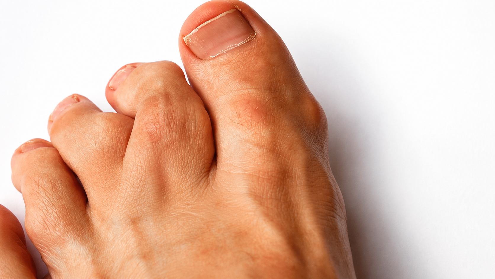
Ollier Disease is a rare skeletal disorder characterized by the presence of multiple enchondromas, which are benign cartilage tumors located within the bones. These growths usually appear near the bone growth areas, especially in the long bones of arms and legs, and can also affect hands, feet, pelvis, ribs, skull, or spine.
Unlike an isolated enchondroma, which can appear without causing major problems, in Ollier Disease, the enchondromas are multiple, can alter the bone architecture and cause deformities, limb shortening, pathological fractures, and significant differences in arm or leg length. It is a rare disease, estimated at approximately 1 case per 100,000 people, and usually manifests during childhood, although in some cases, signs can be detected from birth.
SYMPTOMS
The 3 most common and representative symptoms of Ollier Disease are:
Progressive bone deformity: This is one of the most characteristic signs. As the child grows, the enchondromas can alter normal bone development, causing curvatures, enlargements, asymmetries, or visible changes in hands, feet, legs, or arms.
Difference in limb length: When enchondromas affect the growth plates, they can prevent a bone from growing at the same rate as its counterpart. This can cause limping, gait alterations, joint overload, and body compensations that affect posture and mobility. It is a biomechanical alteration that can change the way the body moves.
Pathological fractures and pain: Bones affected by enchondromas can be more fragile. This means that a fracture can occur with slight blows or efforts that would not normally break a healthy bone. In many cases, pain appears when there is a fracture, advanced deformity, or a lesion that starts to behave aggressively.
A key point is that Ollier Disease is usually asymmetrical. That is, it does not affect both sides of the body equally. It can compromise more a hand, a leg, an arm, or a specific group of bones, generating an irregular pattern that helps the doctor to suspect the diagnosis.
ETIOLOGY, CAUSES, AND DIAGNOSES
Its three main causes or mechanisms are:
Somatic mutations during development: Ollier Disease is not usually hereditary. In most cases, it is associated with mutations that occur after fertilization, during embryonic development. Therefore, not all body cells have the mutation, but only a part of them, a phenomenon known as mosaicism.
Cartilage growth alteration: Enchondromas appear when cartilage remnants remain inside the bone and continue to grow abnormally. This cartilage can interfere with the normal formation of bone tissue, especially in the metaphysis and diaphysis of long bones, key areas for growth during childhood and adolescence.
Benign tumor transformation with malignant risk: Although enchondromas are benign, their multiple presence increases the risk of some lesion changing its behavior. The most important warning sign is the appearance of new pain, rapid growth of a mass, progressive bone destruction, or suspicious changes in images.
Some forms of diagnosis are divided into:
Simple X-rays: These are the first study used. They allow observing lesions within the bone, radiolucent areas, deformities, bone expansion, and characteristic cartilage calcifications.
Magnetic resonance imaging: It is useful to evaluate the extent of the lesions, differentiate benign lesions from suspicious lesions, and analyze soft tissues, bone marrow, and areas with persistent pain.
Computed tomography: Helps to better define the bone structure, especially when there are complex deformities, fractures, or the need for surgical planning.
Orthopedic evaluation and periodic follow-up: The diagnosis is not based only on an isolated image. It is necessary to assess growth, pain, gait, limb length, deformity, fracture risk, and evolution of the lesions over time.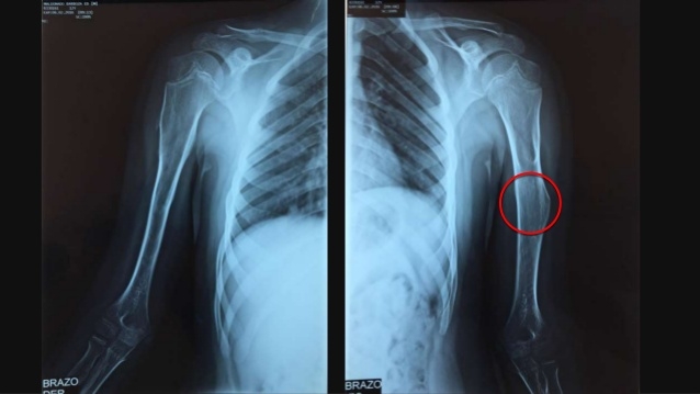
TREATMENTS
There are many strategies, but here we will focus on the 3 most relevant, separating the management of surveillance, orthopedic management, and surgical treatment.
Medical surveillance and control by imaging:
Currently, there is no medication that cures Ollier Disease or makes enchondromas disappear. Therefore, clinical and radiological follow-up is essential. The goal is to detect changes in the size of the lesions, the appearance of pain, progressive deformities, fractures, or signs of malignant transformation.
Orthopedic and functional management:
When there is a difference in limb length, gait alteration, muscle weakness, or postural compensations, the treatment may include insoles, supports, physiotherapy, strengthening exercises, and strategies to improve mobility. Here the goal is not to "cure" the enchondroma, but to reduce the functional impact of the deformity and help the patient move more safely.
Surgical treatment:
Surgery is reserved for specific complications: pathological fractures, significant deformities, persistent pain, severe discrepancy in limb length, or suspicion of malignancy.
And if you or someone close faces physical limitations derived from bone alterations, joint pain, gait problems, or rehabilitation processes, remember that at Pura+ you will find supports, insoles, elastic bands, immobilizers, and physiotherapy products designed to accompany movement, recovery, and daily well-being.
CONCLUSIONS AND REFLECTIONS
Ollier Disease is a rare pathology but deeply important within the world of orthopedics because it demonstrates how a cartilage alteration can modify the entire architecture of the skeleton. It is not just about "deformed bones" or "benign tumors without importance." It is a condition that can affect growth, gait, body symmetry, bone strength, and, in some cases, increase the risk of bone cancer.
Ollier Disease reminds us of something powerful: the human body grows with impressive precision, but when that precision fails, even cartilage, that flexible tissue that normally helps form and protect structures, can become a disordered architect within the bone.
It is not a sentence, but a condition that demands vigilance. Because when cartilage grows where it shouldn't, the skeleton starts to tell a story that needs to be heard in time

The brands Beybies, Pura+ and NrgyBlast belong to Avimex de Colombia SAS. All products have quality certifications and current sanitary registrations and are manufactured under the strictest international standards. To purchase our products, you can access our Shop-On Line. All purchases are backed by a 100% satisfaction or refund guarantee.


